Fibrocystic change
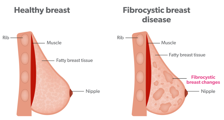
Fibrocystic change is the most common cause of breast lumps in women aged 30–50. Changes occur because some of the breast tissue overreacts to normal monthly changes in hormonal levels. This can lead to scarring around the ducts, which can block the duct and cause tiny cysts to build up. The lumps feel smooth and mobile. Breast tenderness or aching is common, this may be generalised or felt particularly in the upper, outer area of the breasts (near the armpit).
Lumps may fluctuate in size during the menstrual cycle and in some cases, there may be a green or brown nipple discharge which occurs spontaneously (without squeezing).
If the discomfort is mild, no treatment is required for fibrocystic breast changes. Moderate discomfort can be managed by wearing a properly fitted bra and pain medications, like paracetamol. Occasionally, larger cysts causing more severe pain may require aspiration (a doctor uses a needle and syringe to remove the fluid).
Fibrocystic changes in the breast do not increase the risk of developing breast cancer.
Breast cysts
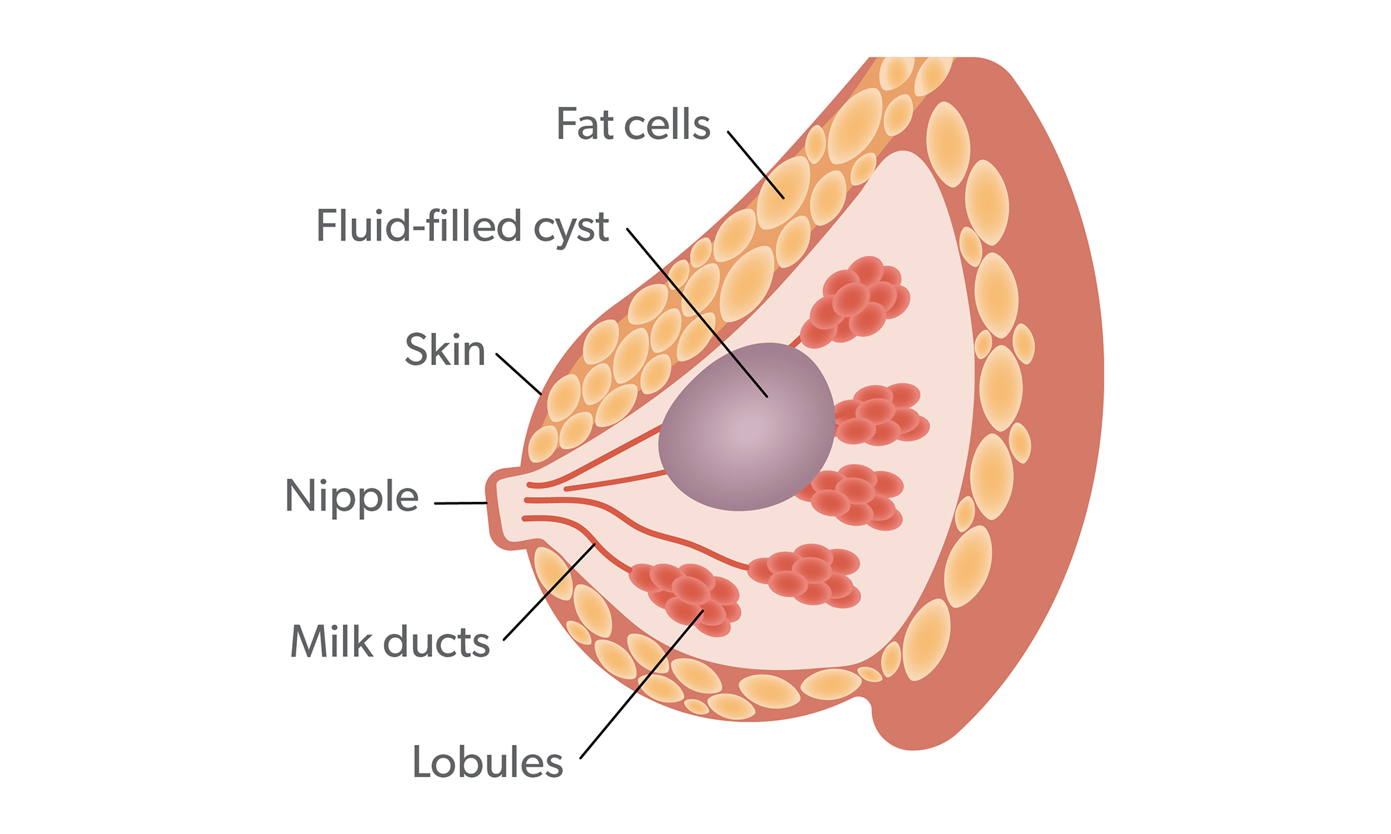
Breast cysts are fluid filled sacs. You may have a single cyst or a number of different sized cysts. Cysts may appear naturally as the breast alters with age, due to normal changes in the oestrogen hormone levels. During the menstrual cycle oestrogen causes fluid to be produced. Although you can develop breast cysts at any age, they’re most common in women over 35.
After the menopause (when your periods stop), as oestrogen levels fall, cysts usually stop forming. Women who have HRT may still get cysts. They may be too small to feel and only detected on imaging or they may be large enough to be felt as a lump. Large cysts may put pressure on other tissue in the breast and cause discomfort.
Cysts can’t be reliably diagnosed on clinical examination alone. Ultrasound will confirm whether the lump is solid or cystic (fluid-filled) and this should be performed before any cyst is drained.
Simple breast cysts are usually left alone. Sometimes the ultrasound will not show the typical appearance of simple breast cysts and a needle biopsy may be necessary to make sure they are breast cysts. If simple breast cysts are very uncomfortable, they may be drained to relieve pain. Occasionally, they can refill or return.
Sebaceous cyst
Sebaceous cysts develop in sebaceous glands which secrete sebum, an oil which coats hair and skin. They are often seen on the face, back and neck but also occur quite commonly in breast skin. The cysts are often surgically removed for cosmetic reasons and because they tend to reoccur and become infected.
Hamartoma
This is a painless soft lump which results from an overgrowth of fibrous, glandular and fatty tissue within a thin capsule of connective tissue. In some cases, it can lead to breast enlargement without a localised lump being felt. It is usually detected on mammogram but requires a biopsy to confirm the diagnosis. Not to be confused with a haematoma which is a collection of clotted blood within the tissues.
Lipoma
Fat necrosis
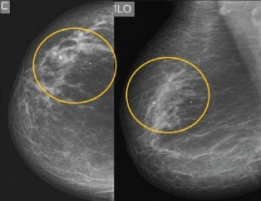
This results from damage to fat cells in the breast, usually caused by trauma, such as seat belt injury after a car crash, surgery, radiation therapy or being on blood thinning medication.
The blood supply to the fat cells is disrupted causing cell damage or cell death. It may be felt as a lump or be seen on a mammogram or ultrasound scan. Sometimes cysts, containing oil, may develop within an area of fat necrosis.
Fat necrosis can look like cancer on a mammogram, so the doctor may want to take a sample (biopsy) to be certain. Usually no treatment is required and the fat necrosis resolves on its own.
Fibroadenomas
Fibroadenomas usually present as a lump or a change detected on ultrasound/mammogram. Fibroadenomas can be different in size, with some women having more than one. The only way to know you have a Fibroadenoma is to have a sample taken (biopsy).
Fibroadenomas are common and thought to be related to the female hormone estrogen. Hormone levels are higher when we are young, pregnant, breast feeding or taking hormone replacement. Although they can appear at any age, they are more likely when aged 15-40 years.
Fibromas are found in the breast lobule, which is the part of the breast that produces milk. Lobules are made up of glands (that hold the milk), ducts (where the milk travels out of the breast) and fibrous tissue (that holds the breast shape). Fibroadenomas occurs when are tissue in the lobule overgrows and becomes hard.
Most will shrink over time, so are usually left alone. If your Fibroadenoma becomes larger or is painful you can speak to a breast specialist about having it removed.